Marjolins ulcer is a variant of skin cancer , it is a ulcerating squamous cell carcinoma . Described by Jean Nicolas Marjolin in 1828.
Which develops in scars it can be burn scar ,osteomyelitis ,venous ulcers, post radiotherapy , pressure sores or post traumatic scars. Unstable wound which are prone to breakdown are frequent site of this cancer . It can be very aggressive tumour . Initial lymphatic spread is slow because of scarring ,but once barrier is breached spread can be aggressive .
Any wound which is not healing for 3 months should be biopsied .
 |
| original ulcer |
30 year old male history of burns sustained burns 20 years ago which was treated conservatively with right get skin grafted . Patient developed ulceration over medial aspect of ankle which refused to heal .
Patient was subjected to biopsy which came out to be squamous cell carcinoma . Surgical Oncology advice was taken and patient was taken for surgery. Lymph node dissection of groin and popliteal region was done . Which came out as negative in frozen section.
 |
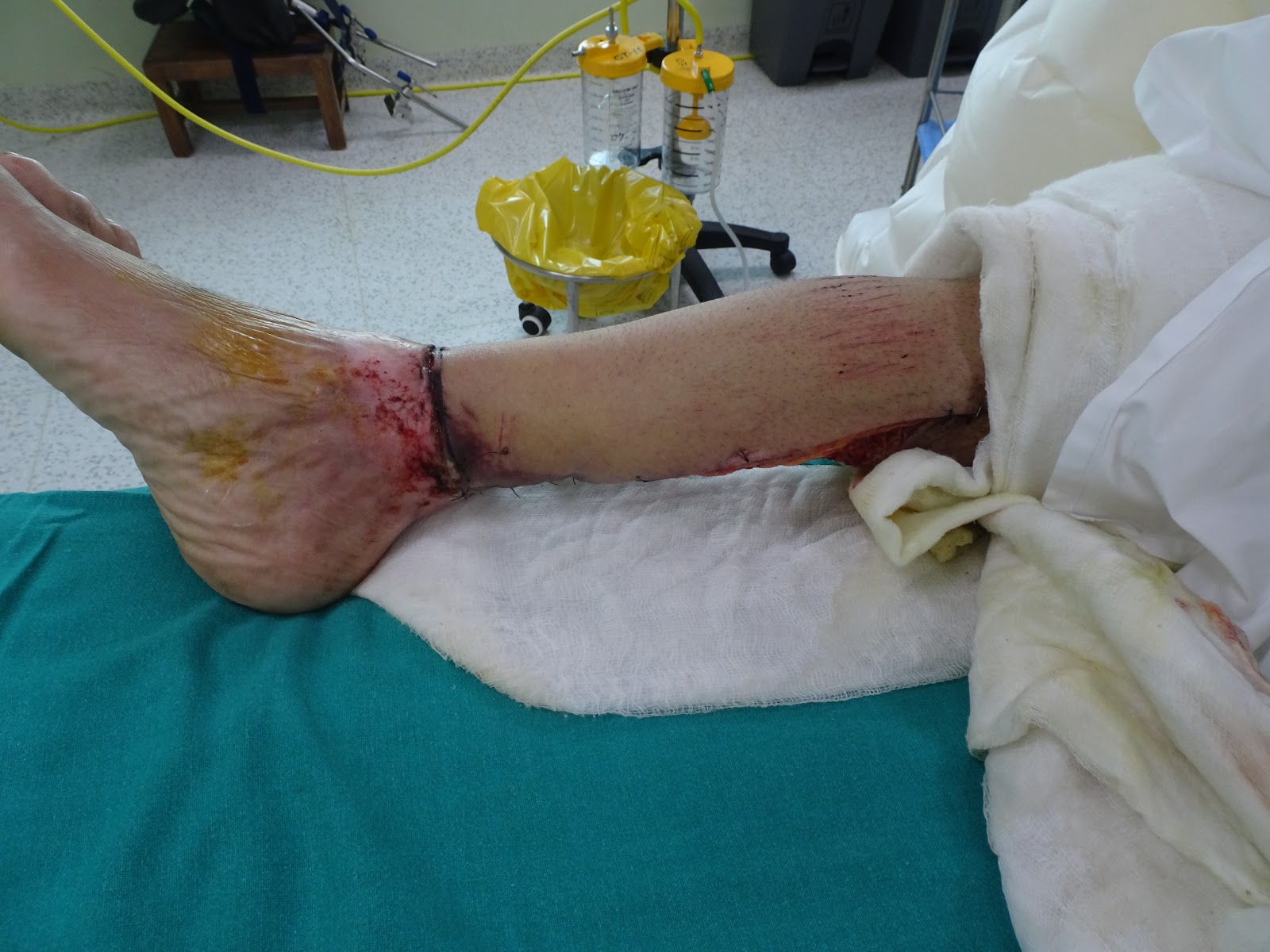
| post op day 5 on the day of skin grafting |
 |
| popliteal fossa wound |
 |
| post op 5 skin grafting the posterior part |
 |
| Onco surgery team removing team |
 |
| final defect and dissection of posterior tibial vessels |
 |
| ALT flap being dissection |
ALT flap of dimension 15 by 15 cm was dissected out and transferred to leg .flap was insetted ,flap artery was anastomosed to end to side to posterior tibial vessels . Both veins were anastomosed , one to saphenous vein and other to veinae comitant . After 5 days patient was again taken up and skin grafting was done. Next day patient was discharged.
Dr Adhishwar Sharma
MB,BS , MS General surgery PGIMER
Mch Plastic Surgery
Fellowship in microvascular surgery
brahmanandclinic.com
 30 year old male history of burns sustained burns 20 years ago which was treated conservatively with right get skin grafted . Patient developed ulceration over medial aspect of ankle which refused to heal .


Comments
Post a Comment